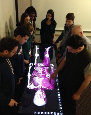
UConn medical and dental students have a new high-tech learning tool. The Anatomage is a virtual anatomy table that works like a giant iPad. The table complements what the students learn with a human cadaver and better prepares them for the future.
Watch the video: http://youtu.be/ylc0cyhS79U
“When the students leave the gross anatomy lab they’ll rely primarily on medical and dental imaging such as MRI and CT images,” explains John Harrison, associate professor in the Department of Craniofacial Sciences. “Those can be challenging to interpret so this virtual anatomy table is a great way to begin to bridge that gap between what they see in the lab and what they’ll see in the rest of their careers.”
The Anatomage has both a male and female cadaver that have been transformed into 3D renderings that students can rotate, manipulate, and cut into cross sections in virtually any plane that they would like to visualize.
“It allows them to look at the standard configurations they would see in medical imaging, axial, coronal, as well as sagittal sections down the middle,” adds Harrison.
“It’s really cool to see it from different angles and things we don’t get to see when they’re in the body sometimes,” says Andrew Glick, first-year medical student. “I think this will definitely complement what we learn in the lab. It’s awesome.”
Harrison has noticed that the students, many who grew up playing video games and using high-tech gadgets, have no problem mastering the Anatomage.
“They’re technical natives. They’ve grown up with the technology, they’re comfortable with it, and they’re very good at it. So this is a great resource for millennial learners,” adds Harrison.
“I think having a little tech-savviness when operating it is helpful,” says Andrew Emery, first-year dental student. “It makes it easier to learn.”
First-year dental student Leila Fussell says, “It’s more interactive which is what we’re used to. I think it will be a great learning tool for all of us, so I’m excited.”
But Harrison says the virtual cadaver will not replace the real thing.
“We have a wonderful anatomical donation program so our gross anatomy labs are provided by donors who will their bodies for the benefit of our medical and dental students and their future knowledge and their future practice. We think that’s an incredibly important thing for our students to experience.”
The first-year medical and dental students are currently using the Anatomage but Harrison says this is just the beginning. The goal is to purchase more tables and create a much larger virtual anatomy lab that will span not only the first year but all four years of medical school.